


Blog Details
By Dr. Tawfic Swaid
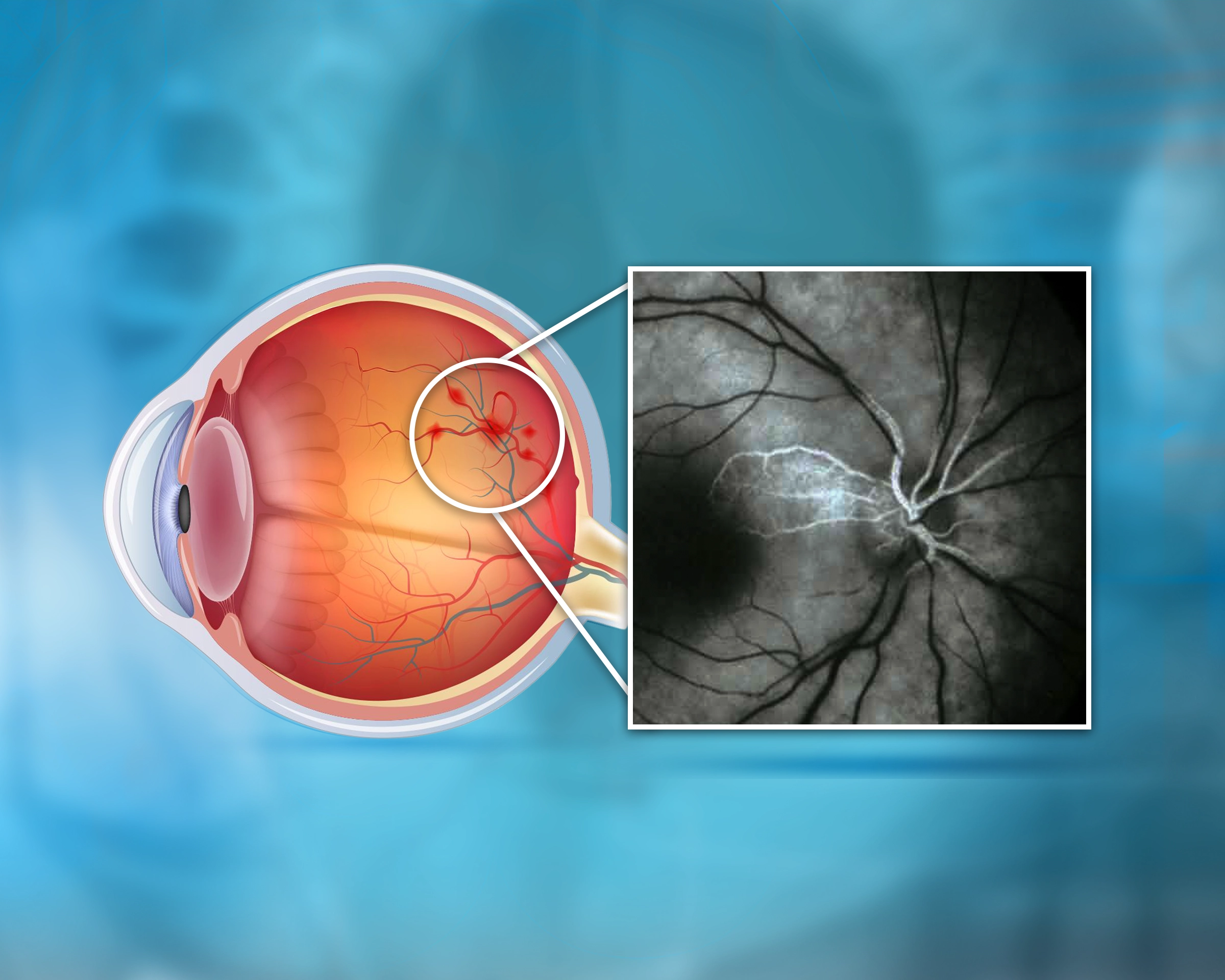
Retinal Artery Occlusion
What Is Retinal Artery Occlusion ?
Retinal Artery Occlusion (RAO) is a sudden blockage of blood flow through the central or branch retinal artery, resulting in acute and painless loss of vision in one eye.
It is considered an ocular stroke, most commonly caused by an embolus or thrombus that obstructs retinal circulation.
The central retinal artery occlusion (CRAO) is the most severe form, while branch retinal artery occlusion (BRAO) affects only part of the retina.
Symptoms of Retinal Artery Occlusion
-
Sudden, painless loss of vision in one eye.
-
Partial vision loss or visual field defect.
-
Blurred or dim vision.
-
Dark or blind spots (scotomas).
-
Occasionally, transient visual loss that recovers briefly (amaurosis fugax).
Causes of Retinal Artery Occlusion
RAO results from interruption of retinal blood flow by :
-
Emboli from the carotid arteries or heart valves.
-
Atherosclerosis causing arterial narrowing.
-
Cardiac disorders (atrial fibrillation, valvular disease, endocarditis, or myxoma).
-
Systemic hypertension, diabetes mellitus, and hyperlipidemia.
-
Temporal arteritis (Giant Cell Arteritis)—an inflammatory cause in older patients.
-
Blood disorders (sickle cell disease, coagulopathies).
-
Oral contraceptives, pregnancy, or intravenous drug use.
Risk Factors of Retinal Artery Occlusion
-
Age > 60 years.
-
Male gender.
-
Smoking.
-
High cholesterol.
-
Hypertension and diabetes.
-
Cardiovascular disease.
-
Carotid artery plaques or narrowing.
Complications of Retinal Artery Occlusion
-
Permanent vision loss.
-
Ischemic optic neuropathy.
-
Neovascularization of the iris or retina.
-
Secondary neovascular glaucoma.
Diagnosis of Retinal Artery Occlusion
-
Comprehensive eye exam and fundoscopy revealing a pale retina with a cherry-red spot.
-
Visual field testing to map vision defects.
-
Fluorescein angiography to visualize blood flow blockage.
-
Optical Coherence Tomography (OCT) for retinal layer evaluation.
-
Systemic work-up: ECG, carotid Doppler, blood tests for glucose, lipids, and clotting status.
Treatment of Retinal Artery Occlusion
RAO is an ophthalmic emergency. Prompt intervention within 90 minutes offers the best chance to restore circulation.
Immediate Management
-
Ocular massage to dislodge the embolus.
-
Inhalation of carbogen (95% O₂ + 5% CO₂) to dilate retinal vessels.
-
Lower intraocular pressure using acetazolamide or topical beta-blockers.
-
Anterior chamber paracentesis in select cases.
Secondary Prevention
-
Manage cardiac and vascular risk factors.
-
Control blood pressure and diabetes.
-
Start antiplatelet therapy (aspirin if indicated).
-
Lifestyle modifications (diet, exercise, smoking cessation).
⚠️ RAO is often a harbinger of stroke or heart disease — systemic evaluation is essential.
Prevention of Retinal Artery Occlusion
-
Regular eye check-ups for high-risk patients.
-
Cardiovascular screening and risk factor control.
-
Healthy lifestyle with balanced nutrition and exercise.
-
Smoking cessation.
-
Strict management of blood sugar and cholesterol levels.

Categories
General Eye Care
LASIK & Vision Correction
Cataract & Lens Implants
Glaucoma
Cornea and its diseases
Retinal diseases and diabetes
Children’s Eye Health
Advanced Technology
Recent Blogs
17/01/2026

12/10/2025
12/10/2025
Popular Tags
Contact Us